Toggle light / dark theme

AI Tool Sets New Standard in Diagnosing Rare Diseases

A new system, which consists of a large LLM and a network of agentic tools, outperformed several other models and human physicians [1].

Too rare to easily diagnose

Rare diseases can be notoriously hard to diagnose. Patients average over 5 years to receive a correct diagnosis, enduring repeated referrals, misdiagnoses, and unnecessary interventions in what is known in rare disease medicine as ‘the diagnostic odyssey’ [2]. These rare diseases, defined as conditions affecting fewer than 1 in 2000 people, collectively impact over 300 million people worldwide. About 7,000 distinct disorders of this type have been identified, with 80% of them being genetic in origin [3].

Convergence of aging- and rejuvenation-related epigenetic alterations on PRC2 targets

Rejuvenation of tissues in physiologically aging mice can be accomplished by long-term partial reprogramming via expression of reprogramming factors (Oct4, Sox2, Klf4, and c-Myc). To investigate the epigenetic determinants of partial reprogramming-mediated rejuvenation, we used whole-genome bisulfite sequencing to carry out unbiased comprehensive profiling of DNA methylation changes in skin from mice subjected to partial reprogramming, as well as young and untreated old controls. We found a striking convergence of age- and rejuvenation-related epigenetic alterations on targets of the Polycomb repressive complex 2 (PRC2), with increased DNA methylation level and entropy over these regions. Native ChIP demonstrated extensive loss of H3K27me3 in aged epidermis compared to young, partially overlapping regions with age- and rejuvenation-related DNA methylation changes. In addition, large H3K9me2-marked “LOCK” heterochromatin domains defined the boundaries for hypomethylated highly entropic regions during aging. These results are also supported by a likewise prominent enrichment of PRC2 targets in gene expression data, suggesting that PRC2 activity can modulate aging and mediate tissue rejuvenation.

Mechanical Dialogues of Life and Death: How External Molecules Entry Triggers a Chromatin‐Cytoskeleton Morphogenetic Duel in Cancer Cells

The next-generation anti-cancer therapeutics must disrupt intracellular mechanics, efficiently eradicating cancer cells, rather than simply intoxicating them. We evaluate the mechanism of action of PCMS, a PAMAM-based supramolecule that eradicates cancer cells by reorganizing their internal mechanics rather than their genes. Once internalized, PCMS self-assembles into a perinuclear ring that severs nucleus-cytoskeleton communication. We observed PCMS’s dual-intelligent mechanisms of action: Cytoskeletal rescue, where actin-microtubule filaments move towards the PCMS ring, treating it as a surrogate plasma membrane, attempting to restore vesicular trafficking; Nuclear counter-expansion, where chromatin-lamina condensates undergo stepwise viscoelastic transitions that push the nuclear envelope outward to reestablish membrane contact. These contradictory forces amplify mechanical stress, driving super-critical strain and nuclear lysis without broad transcriptional modulations. By geometry alone, PCMS collapses the actin-microtubule-nucleus continuum and turns the cell’s adaptive machinery into its own executioner. The discovery that life and death decisions can be reprogrammed through spatial conflict establishes a paradigm of mechanical deception, inaugurating a new class of cellular adaptive feedback-targeted mechanotherapeutics that overcome resistance by exploiting the cell’s own morphogenetic logic.

Abstract: A therapeutic target for terminating the long-lived autoimmune response!

Maureen A. Su find the epigenetic regulator UTX complexes with transcription factors TCF1 and STAT3 to promote pathogenicity of long-lived, stem-like progenitor T cells in models of type 1 diabetes (T1D)


1Department of Microbiology, Immunology, and Molecular Genetics, UCLA David Geffen School of Medicine, Los Angeles, California, USA.

2Department of Pediatrics and.

3Department of Genetics, University of North Carolina at Chapel Hill, Chapel Hill, North Carolina, USA.

DOT1L provides transcriptional memory through PRC1.1 antagonism

Genetic off switch for cancer face_with_colon_three #cancer #cure


Neville, Ferguson et al. show that non-canonical Polycomb repressive complex 1.1-mediated gene silencing is antagonized by DOT1L and is required for the therapeutic efficacy of Menin and DOT1L inhibitors in mixed-lineage leukaemia.

Identifying aggressive prostate cancer with multi-omics

Prostate cancer often develops very slowly. For the vast majority, this is a disease that you live well with, without the need for treatment, but some get an aggressive variant with recurrence of cancer even after surgery. The disease behaves very differently from patient to patient. Understanding what makes the cancer aggressive is crucial for better diagnostics and treatment, says the author.

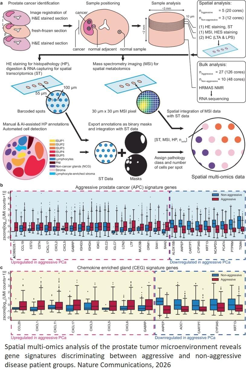
Aggressive cancer has its own gene expression: The researchers identified a pattern in the gene expression of the tumor itself in prostate tissue in patients with a high risk of recurrence and spread. This signature can become a new tool for distinguishing between patients who need intensive care and those who can manage with less intensive follow-up.

Inflammation of apparently healthy tissue: Signs of inflammation and changes in metabolic processes were also found in the normal tissue close to the cancerous tumor. These glands had high activity of neurotransmitters that attract immune cells, and an increased occurrence of a cell type that can trigger inflammatory reactions. At the same time, the levels of important substances had decreased, suggesting that the gland had lost its normal function.

“Aggressive prostate cancer appears to be associated with inflammation in the area around the cancer cells, combined with specific genetic signatures and metabolic changes in the prostate tissue. This knowledge can provide better methods for early identification of patients at high risk,” says the author. ScienceMission sciencenewshighlights.


The research lays a foundation for the possibility that aggressive prostate cancer can probably be detected through a few drops of semen or blood in the long term.

Prostate cancer is the most common form of cancer among men in Western countries.

Germline mutations in DICER1 and DGCR8 can lead to a range of thyroid conditions

Here, Barbara Rivera & team report on the benign-to-malignant progression route in DICER1/DGCR8-thyroid lesions, identifying a DICER1-cancer epi-signature using multi-omic profiling:

The image depicts a thyroid lesion from a sporadic DICER1 case with immunofluorescent staining for pan-cytokeratin (green) and vimentin (red). Enclosed areas represent selected regions of interest.


1Program in Molecular Mechanisms and Experimental Therapy in Oncology (Oncobell), Bellvitge Biomedical Research Institute (IDIBELL), L’Hospitalet de Llobregat, Barcelona, Spain.

2Genetics Program, Faculty of Biology, and.

3Department of Biomedical Sciences, Faculty of Medicine and Health Sciences, University of Barcelona, Barcelona, Spain.

Comparative single-cell lineage bias in human and murine hematopoietic stem cells

A comparative single-cell analysis reveals similarities and differences in lineage bias between human and murine hematopoietic stem cells. This work deepens our understanding of how lineage commitment is regulated across species and provides a valuable framework for translating insights from mouse models to human hematopoiesis.


The commitment of hematopoietic stem cells (HSC) to myeloid, erythroid, and lymphoid lineages is influenced by microenvironmental cues, and governed by cell-intrinsic and epigenetic characteristics that are unique to the HSC population. To investigate the nature of lineage commitment bias in human HSC, mitochondrial single-cell assay for transposase-accessible chromatin (ATAC)-sequencing was used to identify somatic mutations in mitochondrial DNA to act as natural genetic barcodes for tracking the ex vivo differentiation potential of HSC to mature cells. Clonal lineages of human CD34+ cells and their mature progeny were normally distributed across the hematopoietic lineage tree without evidence of significant skewing. To investigate commitment bias in vivo, mice were transplanted with limited numbers of long-term HSC (LT-HSC). Variation in the ratio of myeloid and lymphoid cells between donors was suggestive of a skewed output but was not altered by increasing numbers of LT-HSC. These data suggest that the variation in myeloid and lymphoid engraftment is a stochastic process dominated by the irradiated recipient niche with minor contributions from cell-intrinsic lineage biases of LT-HSC.

Hematopoietic stem cells (HSC) are classically considered to have the capacity for complete regeneration of the hematopoietic compartment. More recent analyses indicate additional complexity and heterogeneity in the HSC compartment, with lineage-restricted or lineage-biased HSC considered a feature of mammalian hematopoiesis.1–13 A partial differential equation model to study relationships between hematopoietic stem and progenitor cells (HSPC) emphasizes that myeloid bias cannot be accounted for solely by short-term HSC bias during inflammation but rather involves a combination of HSC and progenitor cell biases.14 Central to the concept of lineage bias is an assumption that cells used for studying HSC commitment are HSC and not multipotent progenitors or lineage-committed progenitors. Changes in differentiation of cells downstream of the long-term HSC (LT-HSC) must also be evaluated when considering the potential lineage bias of a LT-HSC.

Senescent astrocytes discovered in Alzheimer’s brains point to new treatment targets

Researchers from the NeuroAD group (Neuropathology of Alzheimer’s Disease) within the Department of Cell Biology, Genetics and Physiology at the University of Málaga, also affiliated with IBIMA–BIONAND Platform and CIBERNED, have made a pioneering breakthrough in the fight against this disease by identifying astrocytes as a promising cellular target for the development of future therapies.

The study demonstrates, for the first time, the presence of senescent astrocytes—cells that remain alive but have lost their functional capacity—in the brains of Alzheimer’s patients, positioning this cellular aging process as a key mechanism in neurodegeneration.

The research, published in the journal Journal of Neuroinflammation, was led by Dr. Antonia Gutiérrez, Professor of Cell Biology and Principal Investigator of the NeuroAD group, together with Dr. Juan Antonio García León, Associate Professor of Cell Biology. Other contributors to the study include Laura Cáceres, Laura Trujillo, Elba López, Elisabeth Sánchez, and Inés Moreno.

New AI model could cut the costs of developing protein drugs

Industrial yeasts are a powerhouse of protein production, used to manufacture vaccines, biopharmaceuticals, and other useful compounds. In a new study, MIT chemical engineers have harnessed artificial intelligence to optimize the development of new protein manufacturing processes, which could reduce the overall costs of developing and manufacturing these drugs.

Using a large language model (LLM), the MIT team analyzed the genetic code of the industrial yeast Komagataella phaffii — specifically, the codons that it uses. There are multiple possible codons, or three-letter DNA sequences, that can be used to encode a particular amino acid, and the patterns of codon usage are different for every organism.

The new MIT model learned those patterns for K. phaffii and then used them to predict which codons would work best for manufacturing a given protein. This allowed the researchers to boost the efficiency of the yeast’s production of six different proteins, including human growth hormone and a monoclonal antibody used to treat cancer.

/* */